
Figure 6.
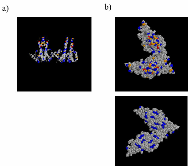
Sequence variation in the context of capsid structure. The crystal structure formed by four HBVc subunits was displayed and coloured using RasMol software. Positions containing 2 polymorphisms are coloured blue, 3 polymorphisms are coloured orange, and 4 or above are coloured red. Grey amino acids are invariant. a) The structure is displayed in ribbon form, showing a vertical section though the capsid, with two spikes projecting upwards, and the internal face of the capsid shown at the bottom of the picture. b) The structure is displayed in space fill. Upper Panel The structure is displayed so that the spikes and outward surface of the capsid are shown towards the viewer, and only the outer surface of the capsid is visible. Lower Panel The structure is rotated by 180 degrees so only the lower (inner) face of the capsid is visible.