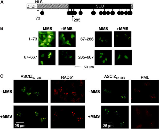
Figure 6.

Identification of a focus-forming ASCIZ core domain. (A) Schematic diagram of ASCIZ domains with arrows indicating truncation points. (B, C) Representative micrographs of focus formation by the indicated ASCIZ fragments in the absence or presence of 0.02% MMS for 4 h in transiently transfected U2OS cells, and co-staining of GFP-ASCIZ residues 67–286 with Rad51 (C, left) and PML antibodies (C, right). Note that micrographs were taken from densely grown fields of cells, and that Rad51 foci are considerably more intense in ASCIZ core domain-expressing cells.
