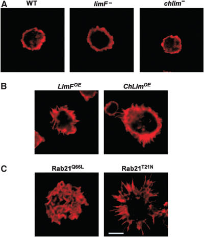
Figure 3.

Actin organization in LimF, ChLim, and Rab21 mutant cells. Various strains of Dictyostelium were grown in shaking suspension, fixed with formaldehyde, stained with rhodamine-labeled phalloidin, and analyzed by confocal microscopy. (A) limF- and chlim-null cells display a cortical ring of F-actin similar to wild type (WT). (B) LimFOE and ChLimOE cells show a modest increase in F-actin-rich filopodia. (C) Cells expressing the constitutively active Rab21Q66L have actin-rich, ruffles over their entire surfaces. Cells expressing the dominant-negative Rab21T21N show enhanced actin microspikes; the bar inset represents 10 μm and is comparable for all panels.
