
Figure 1.
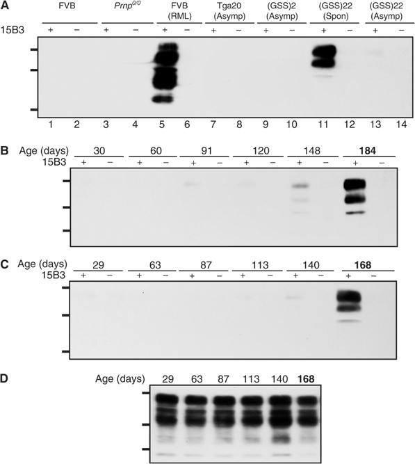
Spontaneous disease in Tg(GSS) mice correlates with the accumulation of 15B3-reactive MoPrP-P101L. (A) Brain extracts were treated with anti-mouse IgM-coupled magnetic beads in the presence or absence of Mab 15B3 as indicated and immunoprecipitates were analyzed by SDS–PAGE and Western blotting as described. Lanes 1 and 2, uninoculated FVB mouse; lanes 3 and 4, Prnp0/0 mouse; lanes 5 and 6, FVB mouse inoculated with mouse-adapted RML scrapie prions; lanes 7 and 8, asymptomatic Tga20 mouse; lanes 9 and 10, asymptomatic Tg(GSS)2 mouse killed at 572 days of age; lanes 11 and 12, spontaneously sick Tg(GSS)22 mouse killed at 188 days of age; lanes 13 and 14, asymptomatic Tg(GSS)22 mouse killed at 30 days of age. (B, C) Kinetics of disease-associated MoPrP-P101L accumulation in Tg(GSS)22 mice. Brain tissues were collected from two independent birth cohorts of Tg(GSS)22 mice killed at various ages, as indicated. The second cohort of Tg(GSS)22 mice was established approximately 1 year after the first. Brain extracts were treated with anti-mouse IgM-coupled magnetic beads in the presence or absence of Mab 15B3 as indicated and immunoprecipitates were analyzed by SDS–PAGE and Western blotting using HRP-conjugated anti-PrP antibody 6H4. (D) Equal amounts of protein in the brains of Tg(GSS)22 mice killed at different ages, as indicated, were analyzed by SDS–PAGE and Western blotting. In (B–D), the ages of asymptomatic Tg(GSS)22 mice are shown in plain text, while mice in which clinical symptoms of disease were manifest at the time of killing are indicated by bold type. The positions of protein molecular weight markers of 35.5, 28.8, and 22.0 kDa from top to bottom are shown.