Figure 3.
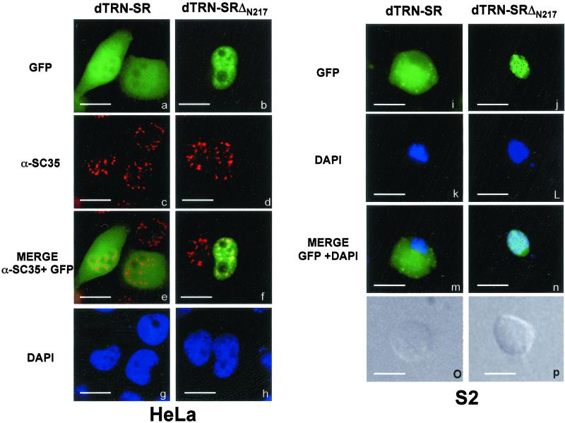
Cellular localization of dTRN-SR GFP fusion proteins in HeLa cells (a–h) and in Drosophila S2 Schneider cells (i–p). Direct fluorescence of GFP-dTRN-SR (a and i) and GFP-dTRN-SRΔN217 (b and j) fusion proteins were analyzed 20 h posttransfection. Expression of fusion proteins was confirmed by immunoblot analysis by using an anti-GFP antibody (our unpublished data). The position of nuclei was confirmed by DAPI staining of the same transfected HeLa (g and h) and S2 (k and l) cells with GFP-dTRN-SR (g and k) or GFP-dTRN-SRΔN217 (h and l). Indirect immunofluorescence staining of HeLa cells in a and b with the αSC35 mAb (c and d, respectively) showed the cellular localization of endogenous SR protein SC35. A merge between a and c (e) and between b and d (f) shows that dTRN-SRΔN217 and SC-35 colocalize in the speckles. The Normarski interference-contrast of the same S2 cells transfected with GFP-dTRN-SR (o) and GFP-dTRN-SRΔN217 (p) are shown. Bar, 10 μm

