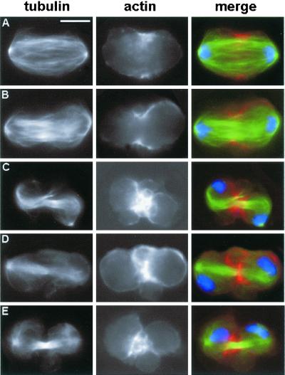
Figure 7.
Abnormal membrane behavior during late telophase of ani (RNAi) cells. Cells were stained for tubulin (green), DNA (blue), and actin (red). (A and B) Anaphase (A) and early telophase (B) figures with normal actin accumulations. (C–E) Late telophases showing large membrane bulges in the cleavage area. The actin-associated fluorescence of these cells has been artificially enhanced to visualize the membrane bulges; these aberrant telophases do not seem to contain more actin than their control counterparts. Bar, 10 μm.

