Figure 3.
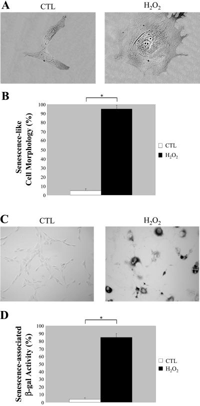
Oxidative stress induces premature senescence in NIH 3T3 cells. NIH 3T3 cells were left untreated or treated with 150 μM H2O2 for 2 h and recovered for 11 d. (A) Cells representative of the two cell populations (untreated or H2O2-treated NIH 3T3 cells) were photographed at a magnification of ×60. (B) Cells were observed under a BX50WI Optical light microscope (Olympus) at a magnification of ×10. The percentage of cells showing a large and flat morphology was scored. Values represent means ± SEM. *P < 0.0005. (C) Untreated and H2O2-treated NIH 3T3 cells were subjected to senescence-associated β-galactosidase activity assay and were observed under a BX50WI Optical light microscope (Olympus) at a magnification of ×10. A representative field is shown. (D) Quantitation of the acid β-galactosidase activity assay shown in C. Note that the treatment with H2O2 successfully promotes premature senescence in NIH 3T3 cells. Values represent means ± SEM. *P < 0.0005.

