Figure 9.
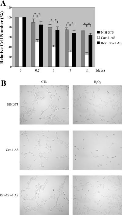
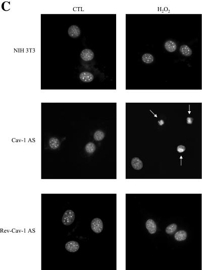
Oxidative stress promotes apoptosis in cells with low caveolin-1 protein expression. (A) Cell death. Normal NIH 3T3 cells, Cav-1-AS cells, and Rev-Cav-1-AS cells were treated with 150 μM H2O2 for 2 h and the cells were allowed to recover for the indicated period of time. The cells remaining in the dish were then collected and counted. Interestingly, H2O2 induces a significantly higher degree of cell death in the Cav-1-AS cells (i.e., that express low levels of caveolin-1). Values represent means ± SEM (n = 8 for each experimental point). *P < 0.001. (B) Cell morphology. Cells were left untreated or treated as in A and allowed to recover for 12 h. Cells were then observed under a BX50WI Optical light microscope (Olympus; ×10 magnification). Note that Cav-1-AS cells clearly show cell shrinkage when stimulated with H2O2. (C) Nuclear morphology. Cells were left untreated or treated as in B, stained with DAPI to visualize their nuclear morphology, and then observed under a Provis fluorescence microscope (Olympus). Note that nuclear condensation is observed only in Cav-1-AS cells.


