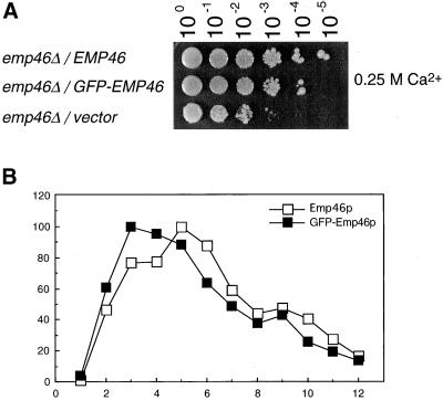
Figure 4.
GFP-3HA-Emp46p is fully functional and shows wild-type subcellular distribution. (A) Cells expressing 3HA-Emp46p (EMP46) or GFP-3HA-Emp46p (GFP-EMP46) were spotted onto a YPD plate containing 0.25 M CaCl2 at 30°C as described in the legend to Figure 2. (B) Sucrose gradient fractionation of GFP-3HA-Emp46p. Whole-cell lysates from cells expressing 3HA-Emp46p or GFP-3HA-Emp46p were separated on sucrose density gradients (20–60%) and fractions were collected from the top. Relative levels of 3HA-Emp46p (□) and GFP-3HA-Emp46p (▪) in each fraction were quantified by densitometry of immunoblots.

