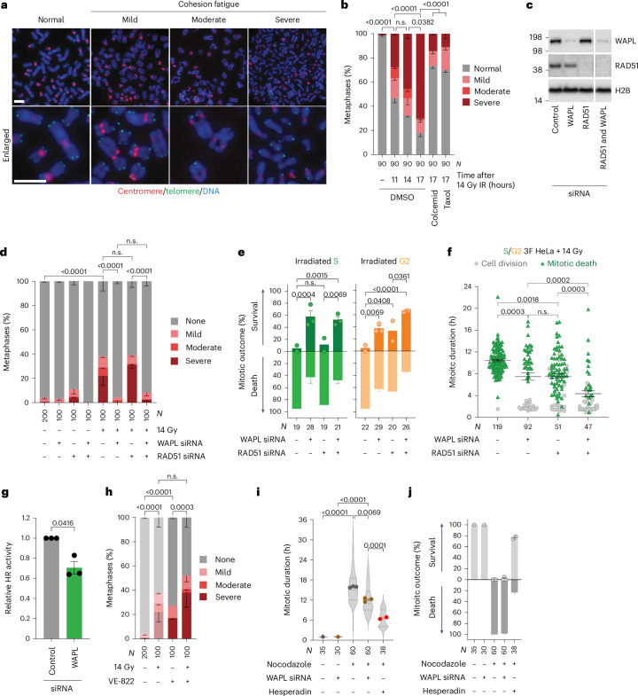
Fig. 5. Cohesion fatigue promotes mitotic death following lethal genomic damage.
a, Cohesion fatigue in HeLa chromosome spreads. Scale bars, 5 µm (representative of n = 5). b, Cohesion fatigue in HeLa ± 14 Gy IR, 100 ng ml−1 colcemid or 5 µg ml−1 taxol (mean ± s.e.m. n = 3, two-sided Fisher’s exact test of N). c, Western blots of HeLa whole-cell extracts (representative of n = 3). d, HeLa cohesion fatigue in the first mitosis after 14 Gy IR (mean ± s.e.m. n = 2, except 0 Gy control siRNA (n = 4), two-sided Fisher’s exact test of N). e,f, First mitosis outcome (e) and mitotic duration (f) in 14-Gy-irradiated asynchronous 3F HeLa (for e, mean ± s.e.m. n = 2, 3, 2 and 3 from left to right, two-sided Fisher’s exact test of N; for f, mean ± s.e.m. and Kruskal–Wallis uncorrected Dunn’s multiple comparisons test of N). g, HR of an I-SceI DSB repair reporter in HeLa (mean ± s.e.m. n = 3, two-tailed paired t-test, t = 4.8, d.f. of 2, 95% confidence interval 0.03 to 0.56). h, Cohesion fatigue in HeLa ± 14 Gy IR and/or 0.2 µM VE-822 (mean ± s.e.m. n = 2, except 0 Gy (n = 4), two-sided Fisher’s exact test of N). The samples without VE-822 are from Fig. 5d. i,j, Mitotic duration (i) and outcome (j) of HeLa ± siRNA, 0.5 µM hesperadin and/or 3.3 µM nocodazole (for i, points means ± s.e.m. of n = 1, 1, 3, 3 and 2 from left to right, violin plot of N, the dotted lines are quartiles and the solid lines medians, Kruskal–Wallis uncorrected Dunn’s multiple comparisons test; for j, mean ± s.e.m. as in i). The control siRNA + nocodazole and/or hesperadin are from Extended Data Fig. 4d. For a–j, N = individual cells/metaphases across all replicates and n = biological replicates. n.s., not significant.

