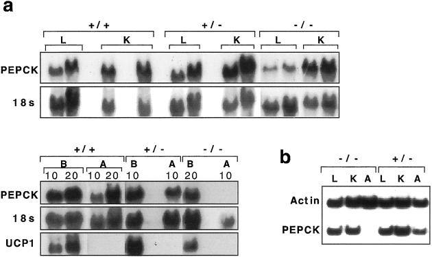
Figure 2.
The expression of PEPCK-C mRNA in wild-type and mutant mice. (a) Northern blot analysis of 15 and 30 μg total RNA from liver (L), kidney (K), and 10 or 20 μg (as indicated above) of brown adipose tissue (B) and white adipose tissue (A) PPARE+/+ (+/+), PPARE+/− (+/−), and PPARE−/− (−/−) mice. The blot was sequentially hybridized to a PEPCK-C cDNA probe, an 18S ribosomal RNA genomic fragment, and a UCP-1 cDNA probe and is representative of five independent experiments. (b) Reverse transcriptase–PCR was performed by using primers for PEPCK-C and β-actin as indicated in Methods. The RNA tested were from Liver (L), Kidney (K), and WAT (A) from PPARE−/− (−/−) and PPARE+/− (+/−) mice.

