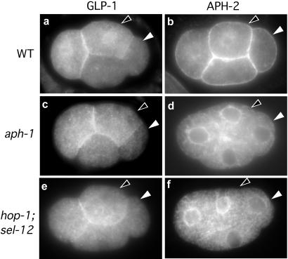
Figure 3.
GLP-1 and APH-2 localization in 4-cell stage embryos. The signaling cell (P2; white arrowhead) and responding cell (ABp; black arrowhead) are indicated in all panels. (a) Immunostaining of GLP-1 in wild type. GLP-1 is expressed on the surface of the anterior-most cell (Left) and its sister (black arrowhead). (b) Immunostaining of APH-2 in wild type. APH-2 is visible on the surface membranes of all four cells. (c and d) aph-1(zu123) embryos labeled as above. (e and f) Embryos deficient in presenilins [sel-12(ty11); hop-1(RNAi)] labeled as above. The level of APH-2 immunostaining is highest when cells are mid-interphase; in the images shown, the GLP-1-expressing cells appear to express a higher level of APH-2 because they are slightly more advanced in the cell cycle than the other two cells.

