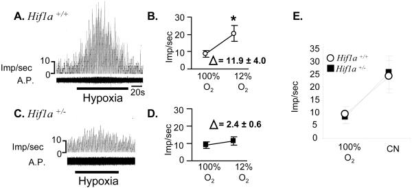
Figure 4.
Response of isolated carotid bodies to hypoxia. Carotid sinus nerve activity was monitored in Hif1a+/+ (A) and Hif1a+/− (C) carotid bodies exposed to 100% O2 before and after exposure to 12% O2 (hypoxia). A.P., action potentials. Mean (± SEM) impulses per second (imp/sec) are plotted and the difference (Δ) between the mean values for 100% and 12% O2 is indicated (B, D). *, P < 0.01 (paired t test). (E) Response of carotid bodies superfused with 100% O2 in the absence or presence of cyanide (CN).

