Abstract
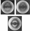
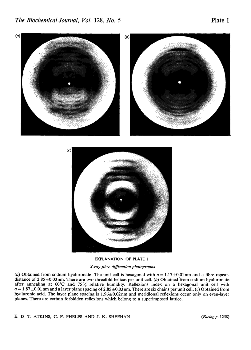
X-ray-diffraction patterns of hyaluronate fibres from a variety of sources were obtained. Sodium hyaluronate gives well-defined patterns which index on a hexagonal unit cell with dimensions a=1.17±nm and a fibre repeat-distance of 2.85±0.03nm. A further form of sodium hyaluronate is produced by annealing at 60°C in 75% relative humidity. This stable state indexes on a hexagonal unit cell of unchanged fibre repeat-distance but with a=1.87nm. The chain conformation is a threefold helix. Analysis of these diffraction patterns led to two tentative structures for sodium hyaluronate, involving different packing of the polysaccharide chains. The significance of side-chain interaction is discussed. Hyaluronic acid produces an X-ray pattern different from that obtained with the sodium salt. The fibre repeat-distance is 1.96±0.02nm and the unit cell appears to be monoclinic. The chain conformation is a twofold helix and conformational change between free acid and monovalent salt is discussed. These findings, together with model-building experiments, are interpreted as indicating a highly ordered structure, and the physical properties of hyaluronate solutions with regard to molecular shape and polyelectrolyte behaviour are rationalized.
Full text
PDF
Images in this article
Selected References
These references are in PubMed. This may not be the complete list of references from this article.
- Anderson N. S., Campbell J. W., Harding M. M., Rees D. A., Samuel J. W. X-ray diffraction studies of polysaccharide sulphates: double helix models for k- and l-carrageenans. J Mol Biol. 1969 Oct 14;45(1):85–99. doi: 10.1016/0022-2836(69)90211-3. [DOI] [PubMed] [Google Scholar]
- Atkins E. D., Mackie W., Smolko E. E. Crystalline structures of alginic acids. Nature. 1970 Feb 14;225(5233):626–628. doi: 10.1038/225626a0. [DOI] [PubMed] [Google Scholar]
- Atkins E. D., Sheehan J. K. Structure for hyaluronic acid. Nat New Biol. 1972 Feb 23;235(60):253–254. doi: 10.1038/newbio235253a0. [DOI] [PubMed] [Google Scholar]
- BITTER T., MUIR H. M. A modified uronic acid carbazole reaction. Anal Biochem. 1962 Oct;4:330–334. doi: 10.1016/0003-2697(62)90095-7. [DOI] [PubMed] [Google Scholar]
- Blackwell J. Structure of beta-chitin or parallel chain systems of poly-beta-(1-4)-N-acetyl-D-glucosamine. Biopolymers. 1969;7(3):281–298. doi: 10.1002/bip.1969.360070302. [DOI] [PubMed] [Google Scholar]
- CARLSTROM D. The crystal structure of alpha-chitin (poly-N-acetyl-D-glucosamine). J Biophys Biochem Cytol. 1957 Sep 25;3(5):669–683. doi: 10.1083/jcb.3.5.669. [DOI] [PMC free article] [PubMed] [Google Scholar]
- Fessler J. H., Fessler L. I. Electron microscopic visualization of the polysaccharide hyaluronic acid. Proc Natl Acad Sci U S A. 1966 Jul;56(1):141–147. doi: 10.1073/pnas.56.1.141. [DOI] [PMC free article] [PubMed] [Google Scholar]
- GOOD T. A., BESSMAN S. P. DETERMINATION OF GLUCOSAMINE AND GALACTOSAMINE USING BORATE BUFFERS FOR MODIFICATION OF THE ELSON-MORGAN AND MORGAN-ELSON REACTIONS. Anal Biochem. 1964 Nov;9:253–262. doi: 10.1016/0003-2697(64)90183-6. [DOI] [PubMed] [Google Scholar]
- Gibbs D. A., Merrill E. W., Smith K. A., Balazs E. A. Rheology of hyaluronic acid. Biopolymers. 1968 Jun;6(6):777–791. doi: 10.1002/bip.1968.360060603. [DOI] [PubMed] [Google Scholar]
- Nieduszynski I., Marchessault R. H. Structure of beta-D-(1-->4')xylan hydrate. Nature. 1971 Jul 2;232(5305):46–47. doi: 10.1038/232046a0. [DOI] [PubMed] [Google Scholar]
- SYLVEN B., AMBROSE E. J. Birefingent fibres of hyaluronic acid. Biochim Biophys Acta. 1955 Dec;18(4):587–587. doi: 10.1016/0006-3002(55)90164-5. [DOI] [PubMed] [Google Scholar]


