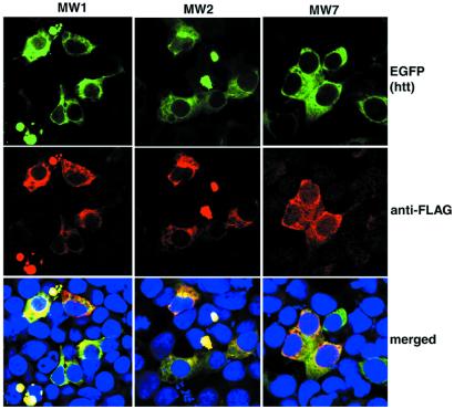
Figure 3.
Anti-htt scFvs colocalize with htt. The 293 cells were cotransfected with mutant htt and an scFv. Twenty-four hours later, cells were fixed and stained with anti-Flag antibody (red), and htt was visualized by the fluorescence associated with its EGFP tag (green). Nuclei were visualized by a DNA stain (blue). In each case, scFvs colocalize with htt.

