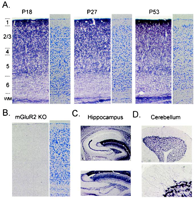
Figure 1.
mGluR2 expression in mouse visual cortex. (A) Upper layers of binocular zone densely labeled across late postnatal development: before (P18), during (P27), or after (P53) the critical period for MD effects (3). Alternate sections, Nissl stain (three mice per age group). (B) No signal in mGluR2 KO mice confirms Ab specificity with no gross change of laminar organization because of deletion (Nissl). (C) Complete absence of mGluR2 in area CA1 of WT mice (Upper). Presynaptic mGluR2 in mossy fiber axons emerging from hippocampal dentate gyrus (Lower). (D) Postsynaptic mGluR2 in somata and dendrites of cerebellar Golgi cells.

