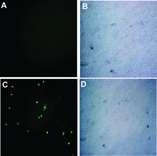
Figure 3.
Chromosomal eBFP conversion after transfection of 293T-eBFP cells with control or 25-LNA-eGFPas ONs. GFP expression was detected by fluorescence microscopy of 293T-eBFP cells 3 days after transfection with control (A) or 25-LNA-eGFPas ONs (C). Brightfield microscopy of 293T-eBFP cells transfected with control (B) or 25-LNA-eGFPas ONs (D). The control ON used was 25-LNA-PDEas of sequence complementary to photoreceptor cGMP-phosphodiesterase. BFP expression could be detected by western blot of 293T-eBFP cells but not by standard fluorescence microscopy due to its low fluorescence yield and strong photobleaching compared with GFP (14).

