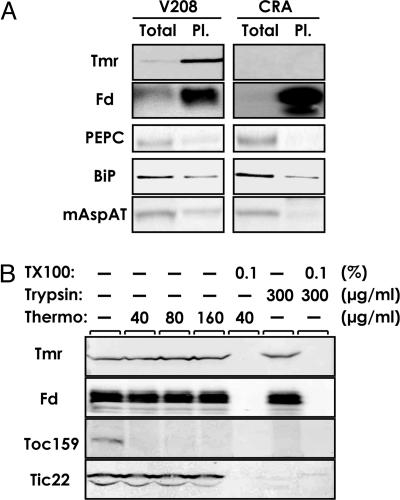
Fig. 5.
Plastid localization of Tmr in crown gall cells. (A) Proteins of total cell extract (Total) and intact plastid fraction (Pl.) (equivalent to 3 μg of protein), prepared from periwinkle V208 or CRA cells, were analyzed by Western blotting. Fd, ferredoxin, a soluble stromal marker; PEPC, phosphoenolpyruvate carboxylase, a cytosolic marker; BiP, binding protein, an endoplasmic reticulum marker; mAspAT, mitochondrial aspartate aminotransferase, a mitochondrial marker. (B) Intact plastids from V208 cells were treated with the indicated concentration of thermolysin (Thermo) on the ice or trypsin (Trypsin) at 25°C for 30 min with or without 0.1% Triton X-100 (TX100). Total proteins from the isolated plastid after respective treatments were then analyzed by Western blotting. Toc159 and Tic22, plastid outer and inner envelope markers, respectively.

