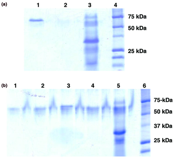
Figure 4.

Tricine SDS-PAGE (T-SDS-PAGE) of proteins purified with the anti-PH-20-antibodies–Sepharose gel. (a) Triton X-100-extracted (lane 1) and octylglucoside-extracted (lane 2) fibroblast cell layers were allowed to interact with the immunoaffinity gel, and fractions eluted with the glycine–HCl buffer were analyzed using T-SDS-PAGE. Lane 3, commercial preparation of bovine testis hyaluronidase; lane 4, Precision Plus Protein standards from Bio-Rad. A strong band was detected in the Triton X-100 extracts, whereas a faint band was barely detected in the octylglucoside extracts. (b) Triton X-100-extracted chondrocyte cell layers and their conditioned media were applied to immunoaffinity columns. Samples eluted with the glycine–HCl buffer were subjected to T-SDS PAGE. Unstimulated chondrocyte cell layers (lane 3; 1.4 μg) and their conditioned medium (lane 1; 0.8 μg). Cell layers of chondrocytes stimulated with 5 ng/ml of IL-1 (lane 4; 1.2 μg) and their conditioned medium (lane 2; 1 μg). Lane 5, commercial preparation of bovine testis PH-20; lane 6, Precision Plus Protein standards from Bio-Rad. Several bands ranging from approximately 60 to approximately 65 kDa can be identified in each specimen.
