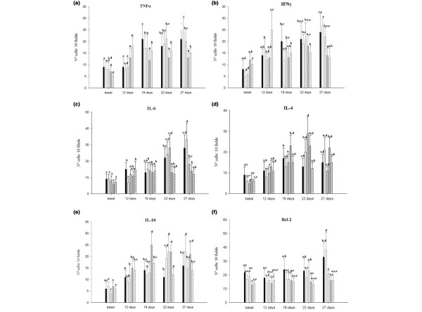
Figure 3.

Cytokine-positive cells in mammary glands. Positive cells for each cytokine were counted in histological sections from mammary glands of the tumour control group (black bars), the P(+) 2d group (white bars), the P(-) 2d group (diagonal lined bars), the P(+) 7d group (grey bars) and the P(-)7d group (horizontal lined bars). (a) Tumour necrosis factor alpha (TNFα), (b) interferon gamma (IFNγ), (c) IL-6, (d) IL-4, (e) IL-10 and (f) Bcl-2. Values are means ± standard deviation for n = 5. Means for each cytokine without a common letter differ significantly (P < 0.05).
