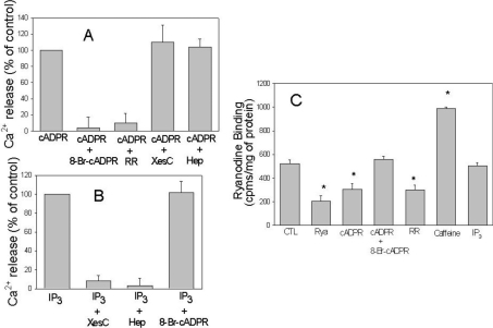
Figure 3. cADPR and IP3 induced Ca2+ release in T. gondii microsomes.
(A) Ca2+ release was induced by 10 μM cADPR in the presence of different inhibitors including 100 μM 8-Br-cADPR, 100 μM Ruthenium Red (RR), 100 μM xestospongin C (XesC) or 1 mg/ml heparin (Hep). (B) Ca2+ release was induced by 10 μM IP3 in the presence or absence of inhibitors as described in (A). In (A, B), Ca2+ release was monitored using the same procedure as that described in Figure 2. (C) Binding of [3H]ryanodine to T. gondii microsomes was measured as described in the Experimental section. Microsomes were incubated for 10 min with 100 μM unlabelled ryanodine (Rya), 10 μM cADPR, 10 μM cADPR with 100 μM 8-Br-cADPR, 100 μM Ruthenium Red (RR), 10 mM caffeine and 10 μM IP3.

