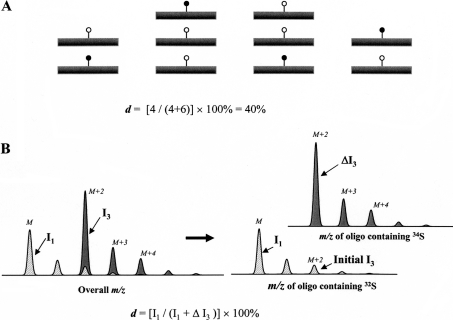
Figure 1. A schematic view of using stable isotope labelling to determine the modification degree at a specific site on a biological molecule.
(A) A modification degree (d) at a specific site on a biological molecule. ●, modification introduced in vivo; ○, stable isotope containing modification introduced in vitro. (B) After a sulphation site is saturated with 34S in vitro, the m/z cluster of an oligosaccharide that carries this site can be considered to be a composite of two. One m/z cluster belongs to the portion of the oligosaccharide generated in vivo (hatched area) and the other belongs to the portion generated in vitro (dark grey area). Intensities of isotopic peaks M and M+2 are I1 and I3 respectively. The enhancement of peak M+2 after the 34S saturation is ΔI3.

