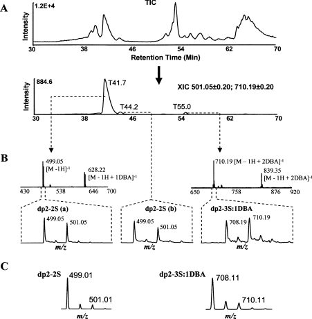
Figure 4. The modification degrees at 6-OST-1 sites.
(A) TIC of the nitrous acid-digested, 34S/6-OST-1-saturated HS sample is followed by the XIC at m/z 501.05±0.2 and 710.20±0.2. (B) The mass spectra and isotope clusters of the three 6-OST-1-labelled oligosaccharides. The three oligosaccharides were identified to be two dp2–2S and a dp2–3S, with the first dp2–2S being the dominant one. Each dp2–2S could form a complex with one DBA and the dp2–3S could form a complex with one or two DBAs. The mass spectrum of the second dp2–2S was similar to that of the first one and was omitted. Sodium adducts were also observed (not labelled). (C) The isotope clusters of dp2–2S and dp2–3S:1DBA, generated by a computer program according to the natural abundances of stable isotopes.

