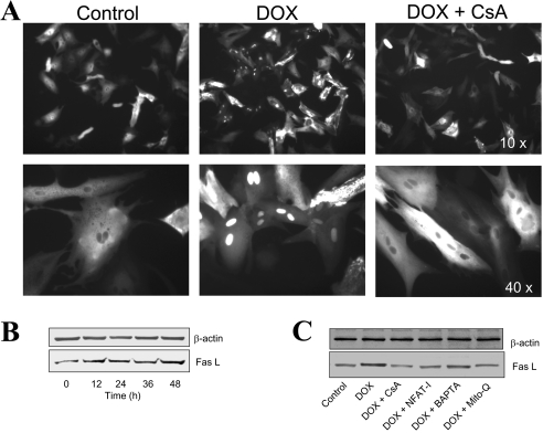
Figure 6. Effect of DOX on NFAT translocation and Fas L expression in primary cultures of cardiomyocytes.
(A) NFAT–GFP-overexpressing cardiomyocytes were treated with 1 μM DOX in the presence and absence of CsA for 16 h, and fluorescence photographs were obtained using a Nikon fluorescence microscope equipped with FITC filter settings. (B) Cardiomyocytes were treated with 1 μM DOX for different time intervals and, after terminating the incubation, cells were collected by gentle scraping, washed thrice with DPBS and lysed in RIPA buffer. Protein (30 μg samples) was resolved on SDS/PAGE (10% gels) and transferred on to a nitrocellulose membrane. Western blot analysis for Fas L and β-actin was performed as described in the Materials and Methods section. (C) Treatment conditions were the same as in (B), except that cells were treated with 1 μM DOX either in the absence or presence of CsA (100 nM), NFAT-I (10 μM), BAPTA (5 μM) or Mito-Q (1 μM). These experiments were repeated three times, and similar results were obtained.

