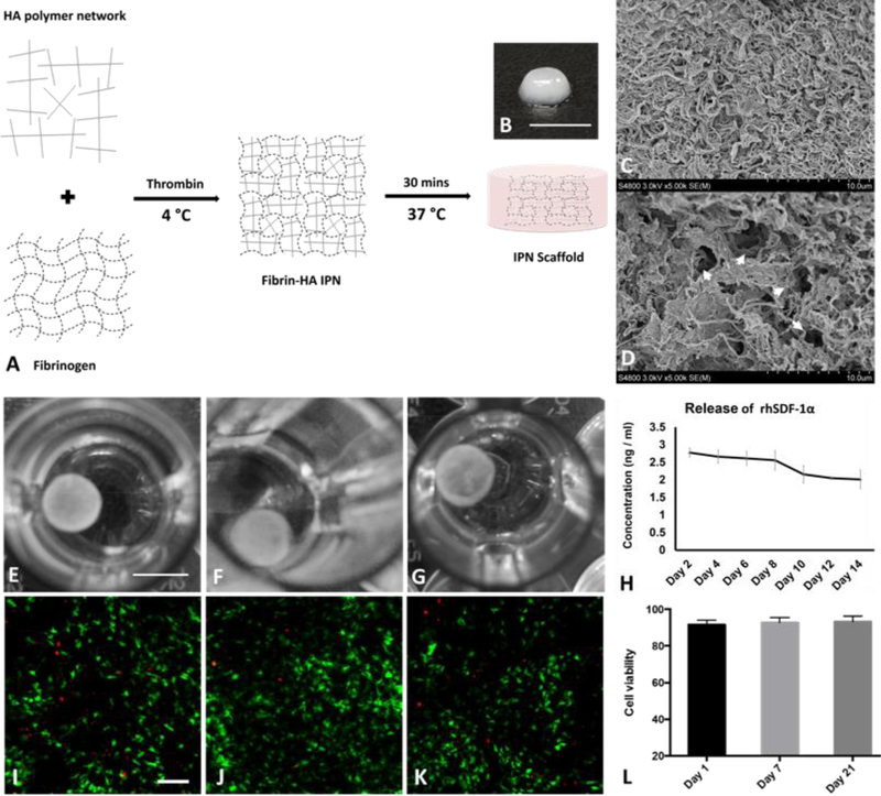
Figure 1.
Fabrication and characterization of IPN hydrogel. (A) A schematic presentation of IPN hydrogel fabrication. Fibrin hydrogel and HA polymer were blended and cross-linked to form interpenetrating polymer network; Macroscopic view of IPN scaffold (B) showed white color and SEM images showed interpenetrated polymer fibers (C) and interconnected pores (D, arrow heads). RhSDF-1α loaded IPN scaffold maintained its integrity in PBS during drug release study for 14 days (E-G); rhSDF-1α protein continued to release from IPN over 14 days (H). Data was presented as mean ± SD (n= 4 for each time point). Encapsulated CPCs were largely viable (green fluorescence) at day 1 (L), 7 (J), (21), with minimal number of dead cells presented (red fluorescence). Average cell viability maintained over 90% for different time points (L). (n= 6 for each time point) Scale bar, B: 5 mm, E-G: 4 mm, and I-K: 500 μm.

