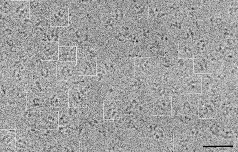
Figure 3.
Typical micrograph of FAS embedded in vitreou ice and recorded at 2.7-μm defocus. The image shown was median-filtered by using a 3 × 3-pixel window to facilitate identification and selection of the molecules. For clarity, several particle images that were selected for image processing have been boxed out. These particles show FAS in different views (e.g. double-arched and dumbbell-shaped). The accompanying close-to-focus micrograph of this area was used to select particles from for image processing. (Bar, 500 Å.)

