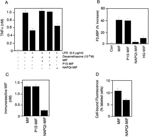
Figure 4.
(A) NAPQI-modified MIF (NAPQI-MIF) shows decreased glucocorticoid-regulating activity on activated monocytes. The ability of various MIF proteins to regulate glucocorticoid suppression of TNFα production in monocytes was assayed as described (35). Monocytes from human peripheral blood were preincubated for with dexamethasone (10−8) or dexamethasone plus MIF (3 nM native MIF, P1S MIF, or NAPQI-MIF) before the addition of 0.5 μg/ml lipopolysaccharide (LPS). NAPQI-MIF was prepared by treatment of MIF with 167 μM NAPQI for 15 min (resulting in 96% inhibition of enzyme activity) and then dialyzed overnight against PBS to remove low molecular weight products. The data shown are mean ± SD of triplicate wells in experiments that were repeated twice. (B) Effect of various MIF proteins on F2,6BP production in differentiated L6 myotubes. L6 rat myoblasts were stimulated with different MIF samples (each at 3 nM) for 24 h, and the F2,6BP then was extracted from cells and measured (15). NAPQI-MIF was prepared by exposing MIF to NAPQI (167 μM) for 5 min. The data shown are mean ± SD and are representative of three independently performed experiments. Con, control. (C) NAPQI-MIF shows decreased immunoreactivity by ELISA. Various MIF proteins (1.4 nM), prepared as described above, were captured with an anti-MIF mAb (10, 13, 17, 21) and quantified by sandwich ELISA (37). (D) NAPQI-MIF shows decreased cell surface binding to human microvascular endothelial cells. Alexa-MIF was formed by reacting MIF with the fluorescent dye Alexa-488. Binding to human microvascular endothelial cells as determined by flow cytometry was compared with Alexa-MIF, which was modified further with NAPQI. The data shown are the mean ± SD of triplicate samples.

