
Figure 3.
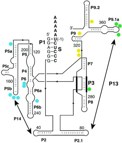
Structural changes in the starting ensemble monitored by DMS accessibility. Residues that increase in protection from DMS in the presence of NaCl are indicated with filled circles. Residues that gave an Na+ concentration for half-maximal protection (K1/2) < 100 mM are colored yellow (C315, C316, C332, C371), residues that gave K1/2 values between 100 mM and 300 mM Na+ are green (C278, A347, A351, A352), and residues with K1/2 > 300 mM Na+ are blue (A151–153, A196, C216, C217, A246). Results are for the standard L-21 ScaI ribozyme in the presence of bound S. The extended version used for single-molecule experiments gave an indistinguishable Na+ dependence of DMS protection (data not shown), suggesting that the structural changes induced by Na+ are similar for the two versions of the ribozyme. The strands that pair to form Alt P3, which can replace P3 (3), are shown with thick lines.