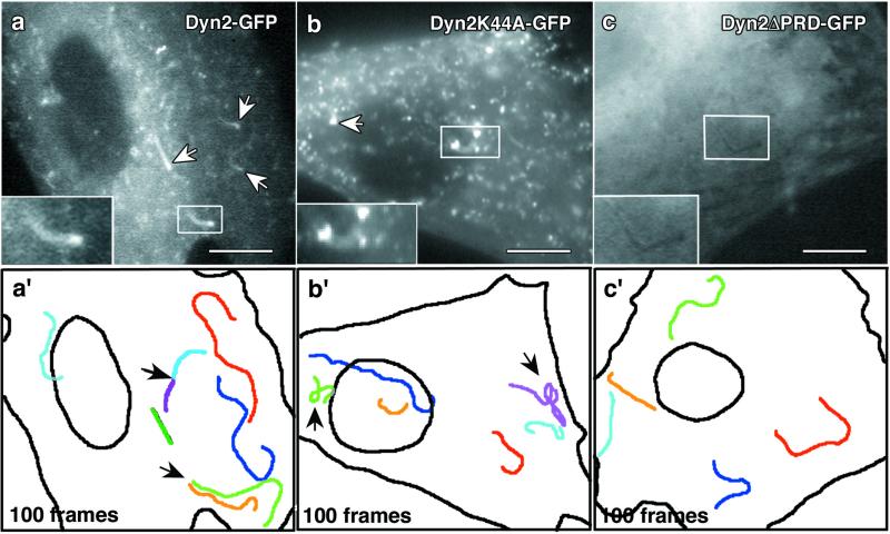
Figure 4.
Mutant dynamin proteins induce abnormal comet movements. Rat fibroblasts coexpressing Myc-PIP5KIα and wild-type or mutant forms of Dyn2-GFP were imaged for 100 frames, and the movement of comet structures was followed. (a) Cells expressing wild-type Dyn2-GFP formed numerous comets (arrows and Inset) that actively incorporated the tagged Dyn2 (see Movie 3). (a′) One hundred frames from the time-lapse images were stacked to show the movement characteristics of individual comets. Each color represents a distinct comet path. Note the linear quality of comet movement. Arrows, multiple comets appeared to form from a single domain. (b) A K44A-GFP-expressing cell with comets (arrows). The comets are fewer and much smaller than those of wild type (see Movie 4). (b′) One hundred stacked frames from the K44A-GFP time-lapse. These comets often had curved and wandering paths (arrows) and were less efficient in their translocation. See the Dyn2K44a-GFP video (Movie 4, arrow on right) for an example of defective movement. (c) Comets in cells expressing the truncated Dyn2ΔPRD-GFP are dark and do not incorporate the mutant protein (see Movie 5). (c′) One hundred frames from the ΔPRD-GFP video revealed that these comets moved in a similar manner to those of wild-type Dyn2, with smooth curvilinear paths. (Bars, 10 μm.)

