Figure 3.
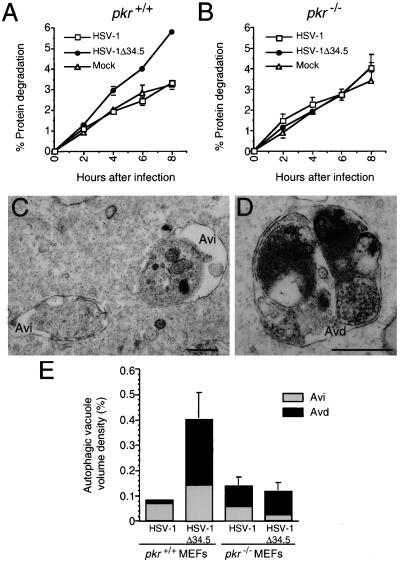
Pkr−/− MEFs are deficient in autophagic protein degradation and autophagic vacuole accumulation induced by α-IFN and HSV-1Δ34.5 infection. (A and B) Cumulative percentage degradation of long-lived cellular proteins in pkr+/+ and pkr−/− MEFs, respectively. Results are mean (± SEM) of triplicate wells. Similar results were obtained in five independent experiments. (C and D) Electron micrographs showing examples of early (Avi) and late (Avd) autophagic vacuoles, respectively, in pkr+/+ MEFs infected with HSV-1Δ34.5. (Scale bars, 0.5 μm.) (E) Volume density of early (Avi) and late (Avd) autophagic vacuoles in pkr−/− and pkr+/+ MEFs infected with wt HSV-1 and HSV-1Δ34.5. Error bars represent SEM of the volume densities of three to five grid squares. Similar results were obtained in two independent experiments.

