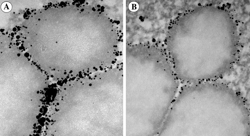
Figure 1.
Peroxisomes in yeast cells were first labeled with the biotinylated Fab fragment antibody against Pex3. This was followed by the second immunolabeling with scFv (A) or Fab (B) against biotin conjugated to NG through MMI linker. Labeling with scFv-NG results in a much higher labeling density than with Fab-NG. Notice that the antigens in the tight interperoxisomal spaces are heavily labeled with the scFv-, but remain unlabeled with Fab-based labels. (Horizontal field width = 685 nm.)

