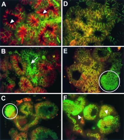
Figure 4.
Loss of Npt2b and NKCC1 expression and cytoplasmic accumulation of β-catenin in Catnb+/Δex3 WAPcre mice at lactation day 1. (A–C) Immunofluorescent staining by using Npt2b (red) and β-catenin (green) primary Abs. (D–F) Immunofluorescent staining using NKCC1 (red) and β-catenin (green) primary Abs. (A) Wild-type alveoli showing apical localization of Npt2b (white arrowhead) and basolateral localization of β-catenin. (B) An alveolus showing loss of apical Npt2b (white arrow) adjacent to alveoli exhibiting apical Npt2b similar to wild-type. (C) Cytoplasmic accumulation of β-catenin (white circle) and loss of detectable apical Npt2b. (D) Wild-type alveoli exhibiting colocalization (orange) of NKCC1 and β-catenin in the basolateral membrane. (E) Loss of alveolar NKCC1 expression (white circle) but normal membranous expression of β-catenin. (F) Loss of alveolar NKCC1 expression (white arrowhead) and cytoplasmic accumulation of β-catenin.

