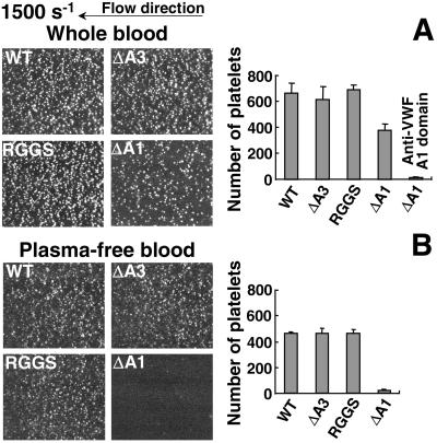
Figure 1.
Interaction of flowing platelets with immobilized recombinant VWF. (A) Whole blood, containing 80 μM PPACK as an anticoagulant, 10 μM PGE1 to block platelet activation, 5 mM EDTA to prevent the function of integrin receptors, and 10 μM mepacrine to render platelets fluorescent, was perfused at the wall shear rate of 1,500 s−1 over recombinant WT-VWF and VWF mutants, as indicated, immobilized on a glass surface. In one set of experiments, the blood also contained 40 μg/ml NMC4 Fab to block the GP Ibα-binding A1 domain of plasma VWF. (B) Plasma-free washed blood cells—containing PGE1, EDTA, and mepacrine, but not PPACK—were perfused instead of whole blood. (Left) Images represent an area of 65,536 μm2, and are single frames from real time recordings taken after 2 min of perfusion on the different surfaces. All interacting platelets exhibited translocation in the direction of flow. The images are representative of four separate experiments for each condition studied, by using blood from different donors. (Right) Bar graphs show the number of platelets interacting with each surface, expressed as mean ± SEM of the four separate experiments.

