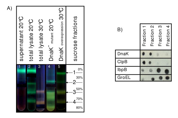
Figure 1.
Separation of recombinant GFP-GST fractions by a sucrose step gradient. A) Distribution of the recombinant protein using cell fractions recovered from different bacterial strains and from bacteria grown at different temperatures. Tube number 1 was loaded with the supernatant separated after lysate ultracentrifugation while total lysates were used for the other experiments. B) Dot-blot for the fractions separated by sucrose step gradient. Each fraction was tested with specific antibodies for the chaperones DnaK, ClpB, IbpB and GroEL.

