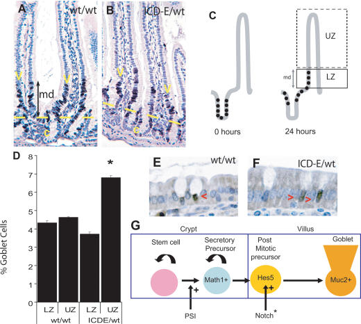
Figure 4.
Notch acts on post-mitotic epithelium. (A,B) BrdU cell migration assays; dark-brown stain indicates BrdU labeling. (A) Ahcre/R26wt/wt. (B) Ahcre/R26ICD-E/wt. (md) Migration distance. (C) Migration assays. BrdU is injected at 0 h, labeling proliferating crypt precursor cells (solid dots); after 24 h these cells and their progeny migrate onto the lower villus, and are detected by immunostaining. (LZ) Labeled zone; (UZ) unlabeled zone. (D) The percentage of Muc2-positive goblet cells in LZ and UZ in Ahcre/R26wt/wt and Ahcre/R26ICD-E/wt mice at 24 h post-induction, based on counts of at least 4000 villus cells in duplicate mice. (*) p = 0.01 by two-tailed t-test compared with LZ in Ahcre/R26ICD-E/wt mice. A typical experiment is shown; error bars indicate standard error of the mean. (E–G) Hes5 immunostaining at 8 h post-induction. (E) Ahcre/R26wt/wt. (F) Ahcre/R26ICD-E/wt. Red arrowheads indicate Hes5-positive cells. (G) Summary of the roles of Notch in the goblet cell lineage: (PSI) presenilin inhibitor; (Notch*) activated Notch.

