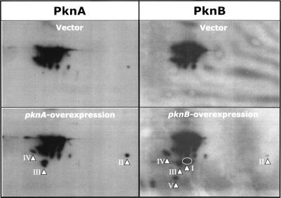
Figure 4.
Comparison by immunoblot analysis of phosphoprotein patterns in whole-cell lysates of M. tuberculosis cells harboring pMH94 vector alone, pMH94-pknA, or pMH94-pknB grown in the presence of inducer. Proteins were prepared from early stationary phase cultures after 24 h of induction, and subjected to 2DGE. Protein was then electrotransferred to PVDF membrane, followed by immunoblotting with a phospho-(S/T)Q-specific antibody and chemiluminescent detection. Arrowheads indicate the spots that showed stronger signals in gels from pknA- or pknB-overexpression cells than the control.

