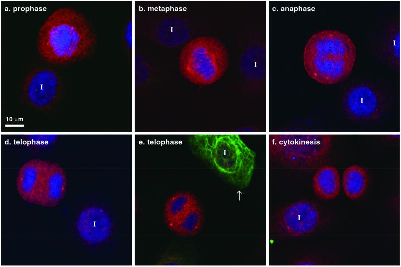
Figure 6.
K5 and K6 LLTPL motif is phosphorylated during mitosis. Primary cultured keratinocytes were triple stained with DL15 (red), toto-3 iodide for DNA (blue), and anti-K8 antibody (green). Most of the cells are K8-negative (except for the cell highlighted by an arrow in e) and K5-positive (unpublished data but similar to Figure 2). DL15 staining increased during mitosis in cells lacking K8. I, interphase cell.

