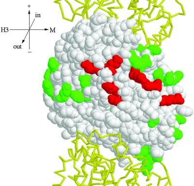
Figure 8.
Two-hybrid interaction footprint of Stu1p on Tub2p. Tub2p is shown as a space-fill model with its GTP-binding site at the top (Richards et al., 2000). The Tub1ps that reside above and below Tub2p in the protofilament are show as backbone traces in yellow. This view shows the outside of the microtubule. Stu1p was tested for interaction with each of the Tub2p mutant proteins by use of the two-hybrid system. Those amino acids whose substitution abolished the interaction are shown in red; those that did not interfere with the interaction are shown in green. Tub2p mutations that abolished interactions with all β-tubulin–binding proteins tested are not shown. Orientation axes: + and − show microtubule orientation; H3 and M show lateral sides marked by the H3 α-helix and M loop, respectively; in and out represent the inside and outside of the microtubule. Structure drawn with RASMOL (Sayle and Milner-White, 1995).

