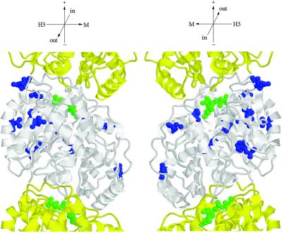
Figure 9.
Location of amino acid substitutions in Tub2p suppressors. Tub2p is shown in white; adjacent Tub1p monomers of the protofilament are shown in yellow (Richards et al., 2000). Amino acids changed in Tub2p suppressors are indicated in blue. GTP is shown in green. The following amino acid substitutions in Tub2p were found to suppress stu1–5: G17C, G17D, W59L, V66F, Q94K, S95R, A102S, V116A, H137N, T149 M, C211F, G223R, F265I, and E288A. C211F was obtained four times; Q94R, S95R, and H137N were each obtained twice; all others were obtained once. Orientation axes: + and − show microtubule orientation; H3 and M show lateral sides marked by the H3 α-helix and M loop, respectively; in and out represent the inside and outside of the microtubule. Structure drawn with RASMOL (Sayle and Milner-White, 1995).

