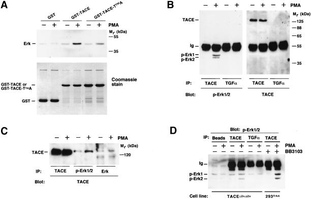
Figure 6.
Association of TACE and Erk. (A) In vitro association of TACE and Erk. GST, GST-TACE, or GST-TACE-T735A coupled to GSH beads was incubated with extracts from control or PMA-treated 293 cells. After incubation at 4°C, the beads were washed with PBS, and Erk associated with the beads was detected by Western blotting with the anti-Erk antibody (top). Bottom, a Coomassie stain of the GST, GST-TACE, or GST-TACE-T735A loaded. (B) 293TrkA cells were treated with PMA or vehicle, and lysates were immunoprecipitated with anti-TACE or anti-TGFα antibodies. Western blots were then probed with anti-Erk (left) or anti-TACE (right) antibodies. (C) 293TrkA cells were treated with PMA or vehicle, and lysates were immunoprecipitated with anti-TACE, anti-p-Erk1/2, or anti-Erk antibodies, followed by Western blotting with anti-TACE antibodies. (D) Active TACE is not required for association with Erk. Extracts from control and PMA-treated TACEΔZn/ΔZn cells were lysed and then protein-A-Sepharose beads were added alone (Beads) or together with the indicated antibodies (anti-TACE or anti-TGFα). In parallel, extracts from 293TrkA cells pretreated with BB3103 (20 μM) were also immunoprecipitated with anti-TACE antibodies. Precipitates were washed and analyzed for Erk presence by Western blotting with anti-p-Erk antibodies.

