
FIGURE 1.
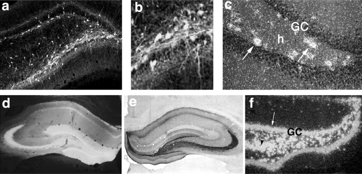
Expression of green fluorescent protein and neuropeptide Y in the hippocampus of representative rats, injected 8 weeks previously with recombinant adeno-associated virus (rAAV) serotype 2 or chimeric serotypes 1 and 2. rAAV serotype-2 induces transgene expression specifically in hilar interneurons with a spread of ∼1.5 mm around the injection site. A, B: Green fluorescent protein in hilar interneurons and their fibers. C: Neuropeptide Y messenger RNA (mRNA) in hilar interneurons (arrows). Note that granule cells do not express the transgene. rAAV chimeric serotypes 1 and 2 induce a larger transgene expression, including hilar interneurons, mossy fibers, and granule cells, with an extension of ∼2.5 mm around the injection site. D: Green fluorescent protein distribution. E, F: Neuropeptide Y immunoreactivity and its mRNA, respectively. Note that intense hybridization signal is observed in granule cells (arrow in F) and CA3 (arrowhead in F). Transgenes were selectively expressed in neurons because they were under control of the neuronal enolase promoter. GC, granule cells; h, hilus. [From Richichi C, En-Ju D. Anticonvulsant and antiepileptogenic effects mediated by adeno-associated virus vector neuropeptide Y expression in the rat hippocampus. J Neurosci (in press), with permission].