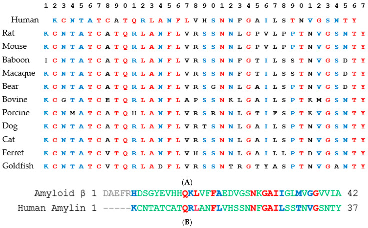
Figure 4.
(A) Multiple sequence alignment of amylin from different species reproduced using the sequences and alignment from Bower and co-workers [40]. (B) Alignment of human amyloid β and human amylin was performed using NCBI COBALT with default parameters. The aligned residues are shown in color (red, blue, and green). Blue shows exact matches, and green shows residues with similar physiochemical features.

