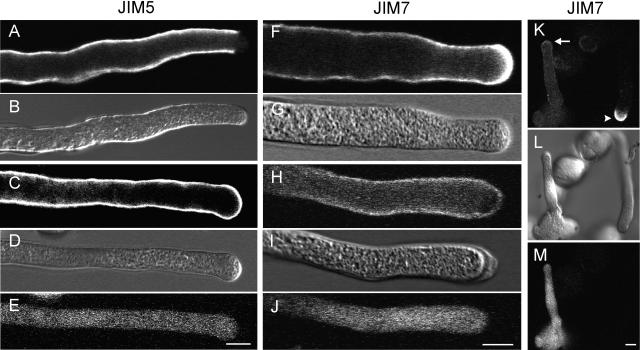
Figure 8.
Comparison of the pectin distribution between transformed pollen tubes expressing the SP-PME-domain (as well as a GFP marker) and nontransformed pollen tubes. DIC images are included as a reference (B, D, G, I, and L), while a GFP signal identifies transformed pollen tubes (E, J, and M). In nontransformed pollen tubes, labeling for JIM5, which detects preferably deesterified pectins, is evenly distributed along the cell wall but absent in the apical region (A). On the contrary, in transformed pollen tubes expressing the PME domain, the JIM5 signal is also present in the apical cell wall region (C). Labeling for JIM7, which detects esterified pectins, is very high in the apical region of nontransformed pollen tubes (F and K arrowhead), while in transformed tubes JIM7 labeling is much weaker at the apical cell wall (H and K arrow). Scale bar = 10 μm.

