
Figure 1.
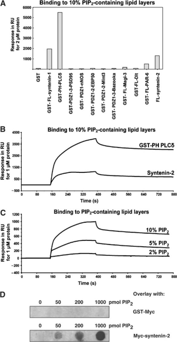
PIP2 binding of selected PDZ proteins. (A) Binding of PDZ proteins or PDZ domains to PIP2 in SPR analysis. Purified recombinant proteins were perfused at 2 μM over a sensorchip coated with vesicles containing 10% PIP2. Values correspond to response units (RU) measured 4 min after perfusion of the proteins. (B) SPR sensograms showing the interaction of syntenin-2 with lipid layers containing 10% PIP2. The syntenin-2 sensogram is compared to the sensogram for GST-PH-PLCδ (positive control for PIP2 binding). The purified recombinant proteins were perfused at 1 μM. (C) SPR sensogram showing that the binding of syntenin-2 to PIP2 increases as the PIP2 concentration in the lipid layers increases. (D) PIP2 binding of syntenin-2 in overlay assay. Increasing amounts of PIP2 (as indicated) were spotted on a nitrocellulose membrane and incubated with GST-Myc as a negative control (upper panel) or Myc-tagged syntenin-2 (lower panel).