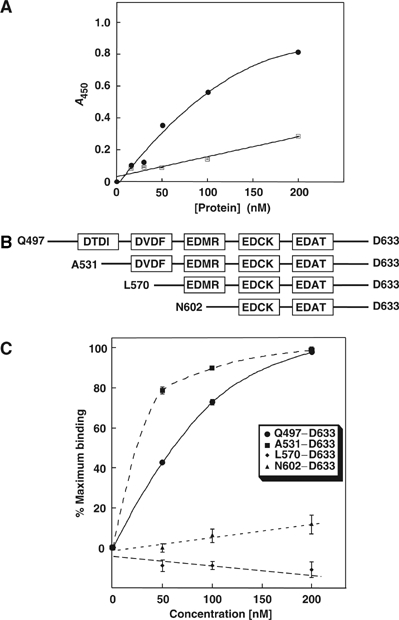
Figure 2.

Identification of the CAII-binding site in the C-terminal region of SLC26A6. (A) CAII, immobilized on a microtiter dish, was incubated with various concentrations of GST (squares) or GSTA6–Q496–D633 (circles). Bound GST, or GST-fusion protein was detected by an enzyme-linked immunosorbant assay. (B) GST fusion proteins correspond to the entire SLC26A6 C-terminus (Q497-D633) and regions progressively truncated from the N-terminus, as indicated in the diagram. Truncation mutants were designed to include different consensus CAII-binding motifs (boxes). (C) Plate-immobilized CAII was incubated with various concentrations of SLC26A6 C-terminal GST-fusion proteins Q497–D633 (•), A531–D633 (▪), L570–D633 (♦) and N602–D633 (▴), and binding was monitored. GST-alone binding has been subtracted.
