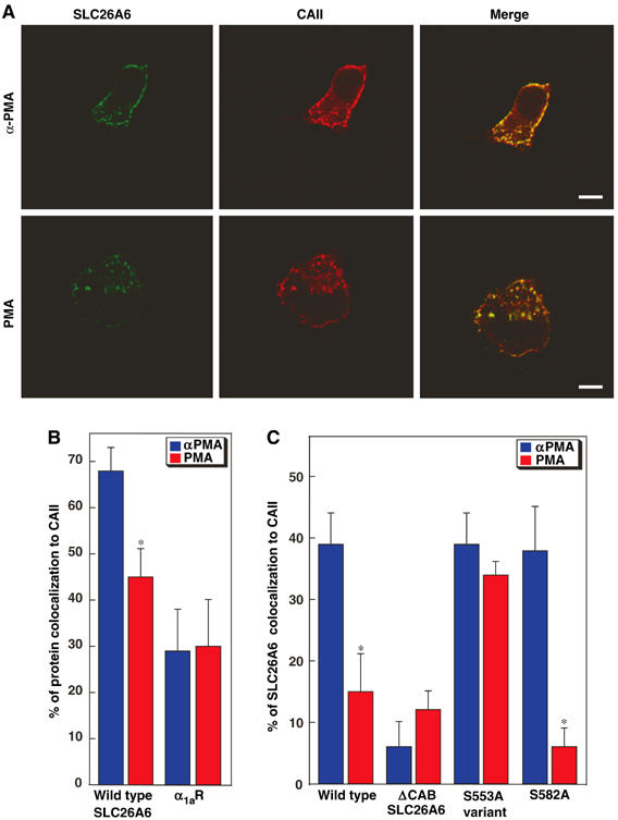
Figure 6.

Effect of PKC activation on CAII cellular localization. HEK293 cells, transfected with an SLC26A6 variant or with the α1a adrenergic receptor (α1aR), were plated on glass slides. Cells were incubated for 1 h with either PMA, or the biologically inactive α-PMA isomer. (A) In WT-SLC26A6, transfected cells were stained with rabbit anti-SLC26A6 antibody, followed by Alexa Fluor 488-conjugated chicken anti-rabbit IgG secondary antibody (SLC26A6, green) or with goat anti-CAII antibody, followed by Alexa Fluor 594-conjugated chicken anti-goat IgG (CAII, red). Colocalization of CAII and SLC26A6 is yellow (merge). Images were collected with a Zeiss LSM 510 laser-scanning confocal microscope. Scale bar=10 μm. (B) Images were analyzed with MetaMorph® Software to quantify the degree of CAII colocalization with either SLC26A6 or α1aR, in cells treated with PMA (red bars) or αPMA (blue bars). *P<0.05 (n=7–20 cells). (C) Colocalization of SLC26A6 variants with endogenous CAII. Values in this panel were corrected for background colocalization represented by the value of α1aR. *P<0.05.
