
Figure 7.
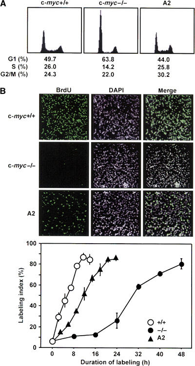
Effect of ATF3 on the cell cycle of c-myc-deficient cells. (A) Asynchronously cycling wild-type, c-myc-deficient, and A2 cells stably expressing ATF3 were analyzed by FACS as in Materials and methods. (B) Each cell line was synchronized by mitotic shake-off in M phase and continuously labeled with 10 μM BrdU as in Materials and methods. Upper panel: Representative data of in situ immunostaining of BrdU-positive cells after 8 h labeling. DAPI denotes nuclear staining. Lower panel: At the indicated time points, BrdU-positive cells were measured by histochemical staining and the percentage of BrdU-positive cells was scored as labeling index. At least 700 cells were scored at each time point.