Fig. 3.
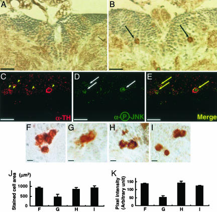
JNK activation in the DM dopaminergic neurons of park1 mutants. (A and B) Frontal sections of the brain were immunostained with rabbit anti-phosphospecific JNK antibody in the brain DM regions of w1118 (A) and park1/park1 (B) and observed under the light microscope. The cells containing phosphorylated and activated JNK were visible only in the park1/park1 brain (black arrows). (Scale bar, 20 μm.) (C–E) The DM region of the 15-day-old park1/park1 brain was costained with sheep anti-TH antibody (C, yellow arrowheads) and rabbit anti-phosphospecific JNK antibody (D, white arrows). (E) Merged image of C and D. (Scale bar, 10 μm.) (F–K) The dopaminergic neurons in the DM regions of 15-day-old flies. (F) parkrv34/parkrv34. (G) park1/park1. (H) UAS-JNKDN/+; Ddc-GAL4/+; park1/park1. (I) hep1/+;park1/park1. TH-positive neuronal cells were marked with rabbit anti-TH antibody, and observed at ×400 under the light microscope. (Scale bar, 5 μm.) The size (J) and color intensity (K) of TH-positive neuronal cells are quantified by using photoshop. Each bar represents mean ± SD from 10 different brains. Dorsal is up in all of the brain sections.

