Fig. 5.
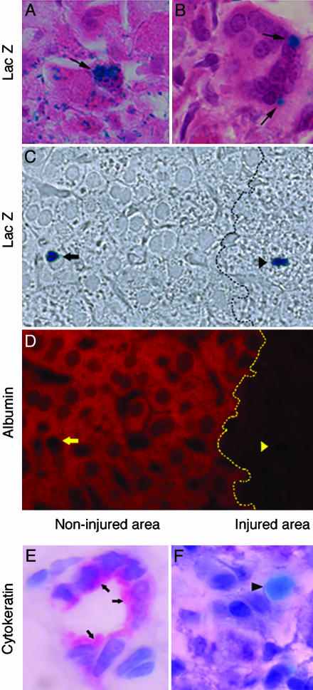
Localization of pancreatic cells within the hepatic lobule. (A) X-Gal staining identifies PDX-1 promoter-driven LacZ expression in cells morphologically similar to hepatocytes near the centrilobular injury (arrow). (B) Some of the stained cells also have an epithelioid appearance, are organized in clusters, and are at the transition between injured zones and intact lobule (arrows). (C) LacZ staining in cells of noninjured (arrow) and injured (arrowhead) areas of the liver lobule without background staining. (D) The same section stained with an anti-albumin antibody (red color) depicting albumin-specific signal in LacZ-stained cells only within noninjured areas (yellow arrow). Dotted black and yellow lines indicate the transition between the noninjured area and injured areas. (E and F) Sections of an intrahepatic bile duct containing epithelial cells stained with anti-cytokeratin antibody (E; arrows), whereas LacZ-stained cells (light blue; arrowhead) are negative for cytokeratin (F). (Magnification: E and F, ×1,000.)

