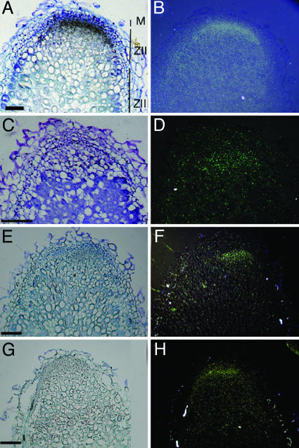
Fig. 1.
In situ localization of DMI2, DMI1, DMI3, and LYK3 mRNA in longitudinal sections of 14-d-old Medicago nodules. (A, C, E, and G) Bright-field images of nodule sections hybridized with 35S-UTP labeled antisense DMI2 (A), DMI1 (C), DMI3 (E), and LYK3 (G) probes. The signal appears as silver grains (black). (B, D, F, and H) Epipolarization images of A, C, E, and G, respectively. M, meristem; ZII, infection zone; ZIII, fixation zone. (Scale bars: 200 μm.)

